These brochures are useful for doctors and optometrists to give out to patients when discussing a condition, or the reason for referring them to see a specialist.
Click to download any of these brochures, or click here to request printed copies for your practice.

Pterygium
A pterygium is a wing-shaped extension of thickened tissue on the surface (conjunctiva) of the white of the eye, which grows onto the adjacent cornea (the window into the eye).
Pterygia are benign growths (not cancers), which can continue to grow across the eye and eventually seriously affect sight. The term is derived from the Greek word “pteru-gion” meaning “little wing”.
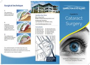
Cataract Surgery
A cataract is gradual yellowing or clouding of the lens inside the eye. In earlier times, cataract surgery was only done when the eyesight was very bad.
Advances in technology mean that modern surgery is less traumatic to the eye. The results are more predictable, there are few side effects and the eye usually recovers quickly.
Modern cataract surgery is done when people find their failing eyesight does not let them do their day to day activities.
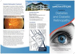
Diabetes and Diabetic Retinopathy
Diabetes can affect the eye in several ways. It can damage your sight by causing cataract, but also more importantly, by causing diabet ic retinopathy.
The retina is the light sensitive film at the back of the eye which changes light into nerve signals that are then transmitted to the brain. Diabetic retinopathy is a potentially blinding complication of diabetes that affects up to a half of diabetics to some degree.
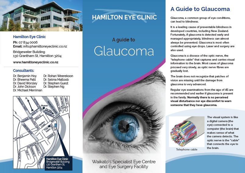
Glaucoma
Glaucoma is a common eye condition that can lead to blindness and is the second most common cause of blindness in New Zealand. Fortunately if glaucoma is detected early and managed appropriately in nearly every case blindness is preventable.
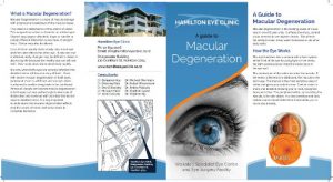
Macular Degeneration
Macular degeneration is the leading cause of visual loss in over 60 year olds. It affects only central vision so there are difficulties with reading, driving and other fine, detailed visual activities.
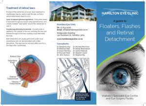
Floaters, Flashes and Retinal Detachment
The vitreous is the gel that fills the eye. Lining the inside wall behind the vitreous is a light sensitive layer we call the retina. As we age the vitreous becomes more fluid-like in some areas, causing clumping of the gel which we occasionally see as floaters in our vision.
Click to download any of these brochures, or click here to request printed copies for your practice.